DUAL- ARM MULTI-MODALITY IMAGING SYSTEM FOR EQUINE APPLICATIONSBASED ON ORIMTECHACE / AVRG TECHNOLOGY
RADIOGRAPHY, TOMOGRAPHY, TOMOSYNTHESIS, AND FLUOROSCOPY IMAGING CAPABILITIES INTEGRATED IN A SINGLE SYSTEM
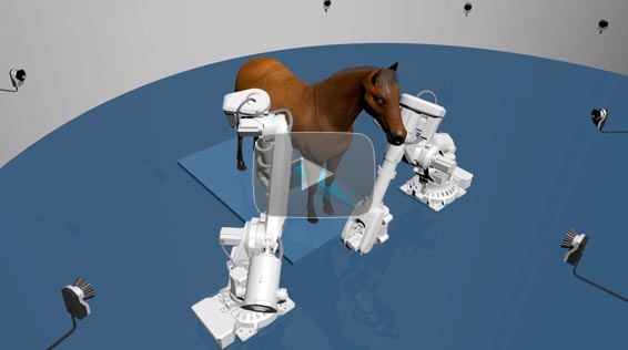
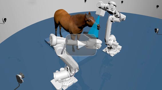
EDAMIS IN LARGE ANIMAL VETERINARY APPLICATIONS: AVOIDING ANESTHESIA DURING TOMOGRAPHIC STUDIES
OF HORSES AND CAMELS
SYSTEM MAIN TECHNICAL SPECIFICATIONS
Common features
- Modalities: Radiography, Fluoroscopy, Cone Beam CT, Tomosynthesis.
- Available X-Ray technique factors are defined by the integrated X-Ray generator, tube, tube assembly, and detector. Currently the supported system configuration are based
on X-Ray Swiss Genesis (Josef Betschart AG, Switzerland) or CPI Indico IQ 100 (Canada ) generator and Varex XRD 4343 RF (Germany) or iRay Mercu1717V (China) panels. COMET generators are also supported. - Collimation: 4 blade PC-controlled automatic collimator
- Acquisition: Orimtech proprietary acquisition/synchronization system
- Object/patient database
- DICOM support; PACS compatible
- Common advantage: high flexibility in selection of patient-system orientation, source-detector distances and system scanning trajectories for all modalities - fully defined by specific robot models.
CBCT specifications
- X-Ray factors, techniques, and frame rate: defined by the integrated X-Ray generator, tube, detector.
- Scanned object positioning/orientation: arbitrary (within the system mechanical accessibility), up to 12 User-predefined scanning trajectory protocols
- FOV adjustment and scouting: collimator light and laser, joystick controlled system motion, linear X-Ray scanning in a single or dual orthogonal planes scout scans to allow for further User-PC interactions to definine area of interest for a scan
- Scan angle: 210 degrees
- Field of view: D240mm x 240mm one slab; with optional 2 stitched slabs with up to D240mm x 420mm of total coverage, depending on tube/detector distance configuration.
- Spatial resolution: 250 micron (mostly defined by a tube focal spot)
- Acquisition technique: pulsed, up to 25 FPS.
- Single slab scanning time: 10-60 sec depending on the X-Ray dosage
- Supported scanning trajectories: circular and non-circular; built-in trajectory registration for the best spatial resolution.
- Ability to scan solid moving objects (motion artifact mitigation): supported by integrated Qualysis visual motion tracking subsystem
- X-Ray technique modulation during the scanning process for the optimal patient dose utilization: supported
- CT reconstruction engine: AVRG with support of scatter, beam-hardening and metal artifact reduction.
- HU accuracy: +/-25 HU on CTP486 section (CATPHAN)
- 3D image viewer: Orimtech MPR proprietary, Fovia HDVR is an option
Fluoro specifications
- X-Ray factors, techniques, and frame rate: defined by the integrated X-Ray generator, tube, detector.
- Automatic brightness control: hardware and software supported
- Real-time display and playback: supported with Orimtech proprietary dynamic contrast enhancement function
- FOV position control: joystick , PC keyboard and touch screen
- Subtractive fluoroscopy techniques (for contrast studies): supported
Radiography specifications
- X-Ray factors, techniques: defined by the integrated X-Ray generator, tube, detector.
- Spatial resolution: mostly defined by X-Ray panel; approximately 5LP for XRD 4343 RF
- Imaged object positioning/orientation: arbitrary, within the system mechanical accessibility
- Source-detector distance: arbitrary, within the system mechanical accessibility
- FOV adjustment: collimator light and laser, joystick controlled system motion.
- DR image processing library: Orimtech proprietary, CVIE (Context Vision)-ready
Tomosynthesis specifications
- X-Ray factors, techniques, and frame rate: defined by the integrated X-Ray generator, tube, detector.
- Scanned object positioning/orientation: arbitrary (within the system mechanical accessibility) , up to 8 User-predefined scanning trajectory protocols
- FOV adjustment and scouting: collimator light and laser, joystick controlled system motion, linear X-Ray scanning in a single or dual orthogonal planes with further User-PC interactions for defining the scanned area
- Scan angle: 15-90 degrees for angular tomosynthesis examinations, 360 degrees for circular motion.
- Field if view: D240mm x 240mm
- Spatial resolution: 200 micron (mostly defined by a tube focal spot)
- Acquisition technique: pulsed, up to 25 FPS.
- Scanning time: 4-20 sec
- Supported scanning trajectories: circular and non-circular; built-in trajectory registration for the best spatial resolution.
- Ability to scan solid moving objects (motion artifact mitigation): supported by integrated Qualysis visual motion tracking subsystem
- X-Ray technique modulation during the scanning process for the optimal patient dose utilization: supported
- Tomosynthesis reconstruction engine: AVRG (Orimtech)
- 3D image viewer: Orimtech proprietary